Human Heredity: Principles and Issues (MindTap Course List)
11th Edition
ISBN: 9781305251052
Author: Michael Cummings
Publisher: Cengage Learning
expand_more
expand_more
format_list_bulleted
Concept explainers
Videos
Textbook Question
Chapter 10, Problem 10QP
- b. Compounds A, B, C, and D are known to be intermediates in the pathway for production of protein E. To determine where the block in protein-E production occurred in each individual, the various intermediates were given to each individual’s cel Is in culture. After a few weeks of growth with the intermediate, the cells were assayed for the production of protein E. The results for each individual’s cells are given in the following table. A plus sign means that protein E was produced after the cells were given the intermediate listed at the top of the column. A minus sign means that the cells still could not produce protein E even after being exposed to the intermediate at the top of the column.
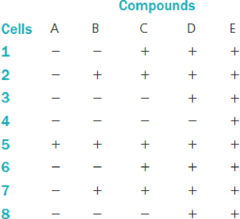
Denote the point in the pathway in which each individual is blocked.
Expert Solution & Answer
Want to see the full answer?
Check out a sample textbook solution
Students have asked these similar questions
A. Lineweaver-Burk plot of the enzyme with increasing amounts of substrate in the absence
or the presence of the inhibitor is shown below.
Graph A : x-intercept
Graph B : x-intercept = - 0.012, y-intercept = 0.8
Graph C : x-intercept = - 0.027, y-intercept = 0.8
Graph D : x-intercept = - 0.039, y-intercept = 0.8
- 0.007, y-intercept = 0.8
Graph A
4
Graph B
Graph C
Graph D
1
-0,04
-0,02
0,00
0,02
0,04
1/[Substrate] (uM)
(i) Which graph indicates an enzymatic reaction without inhibitor?
(ii) Which type of inhibitor is it? Briefly explain.
(iii) Which graph indicates the highest concentration of inhibitor?
(iv) Calculate the Vmax and Km of the graph showing an enzymatic reaction with the lowest
concentration of inhibitor. Show the steps of calculation and unit in your answers.
Keep 2 decimal places in your answers.
1/Rate (umol/min)
a. An oligopeptide ALVGALGATPTPQMWSHSWRGVSIKS was digested with trypsin.Which method would be most appropriate for separating the products: ion exchange or gel filtration chromatography? Explain.b. Suppose that the peptide was digested with cyanogen bromide. What would be the optimal separation technique? Explain
This is an SDS-PAGE gel of the protein insulin. The first lane is the molecular weight standard marker. The second lane NR is the native, non-reduced protein (MW 5.7kDa). The third lane is the protein treated with beta-mercaptoethanol. Please explain what is shown in the NR lane versus the R lane.
Chapter 10 Solutions
Human Heredity: Principles and Issues (MindTap Course List)
Ch. 10.4 - Prob. 1GRCh. 10.4 - Prob. 2GRCh. 10.7 - Prob. 1EGCh. 10.7 - Prob. 2EGCh. 10 - A couple was referred for genetic counseling...Ch. 10 - A couple was referred for genetic counseling...Ch. 10 - A couple was referred for genetic counseling...Ch. 10 - Many individuals with metabolic diseases are...Ch. 10 - Prob. 2QPCh. 10 - Enzymes have all the following characteristics...
Ch. 10 - Questions 4 through 6 refer to the following...Ch. 10 - Questions 4 through 6 refer to the following...Ch. 10 - Prob. 6QPCh. 10 - Prob. 7QPCh. 10 - Prob. 8QPCh. 10 - a. Compounds A, B, C, and D are known to be...Ch. 10 - b. Compounds A, B, C, and D are known to be...Ch. 10 - a. If an individual who is homozygous for the...Ch. 10 - Prob. 12QPCh. 10 - Suppose that in the formation of phenylalanine...Ch. 10 - If phenylalanine was not an essential amino acid,...Ch. 10 - Phenylketonuria and alkaptonuria are both...Ch. 10 - The normal enzyme required for converting sugars...Ch. 10 - Knowing that individuals who are homozygous for...Ch. 10 - Prob. 18QPCh. 10 - A person was found to have very low levels of...Ch. 10 - If an extra nucleotide is inserted in the first...Ch. 10 - Transcriptional regulators are proteins that bind...Ch. 10 - Prob. 22QPCh. 10 - Prob. 23QP
Knowledge Booster


Learn more about
Need a deep-dive on the concept behind this application? Look no further. Learn more about this topic, biology and related others by exploring similar questions and additional content below.Similar questions
- topic: Bradford AssayThere are numerous methods of protein determination in use, but this module focuses on the Bradford assay.The Bradford assay is a dye-binding method that employs Coomassie Brilliant Blue G-250, whose structureis shown in Figure 2.3.4.1. Coomassie Brilliant Blue G-250 is a dye that interacts with proteins throughhydrophobic and electrostatic interactions. What are the identities and functions of the components of the Bradford reagent in protein contentdetermination?arrow_forwardALANINE AMINOTRANSFIRASE AND ASPARTATE AMINOTRANSFERASE (KARMEN МЕТHOD) (C. chemistry) 1. state the reason why substrate must be incubated prior to the addition of serum 2. what part of the steps need extra to avoid positive results? 3. describe the principle of AST/ALT kinetic method.arrow_forwarda. Why proteins are extracted to a buffered solution? Briefly describe the components of ageneralized protein extraction buffer?.b. Describe the basis of affinity chromatography in protein purification.c. What is the most appropriate method of protein elution in affinity chromatography?d. List three examples of commonly employed combinations of ligand and protein in affinitychromatography.arrow_forward
- what is lactose intolerance ? describe the molecular life cycle for this disease. also describe how it occurs in a molecular level detailed mechanism. what causes this disease and how it develops ? provide detailed biochemical phenomena and life cycle for Lactose Intolerance condition.arrow_forward(b) Both laboratories used 10 micrograms of protein each in their kinetic assays. Protein concentrations weredetermined by the Bradford protein assay. Assay conditions employed in the two labs (pH, temperature,etc.) were also identical. What would be the most plausible cause for the discrepancy in the Vmax valuesfor the compound I? Explain.Recall that the Bradford assay measures total protein amounts in sample solution based on complexformation between a dye and proteins. Also, the assay solution used in both labs does not contain anyinhibitors.arrow_forwardSuppose that you are tasked to determine the protein concentration of an unknown protein solution via Bradford assay. You prepared six solutions of bovine serum albumin (BSA) with different concentrations. The initial concentration of the BSA stock solution is 7.50 mg/mL. Approximately 200 µL of Bradford Reagent was added to each of these solutions and the absorbance at 595 nm was taken after 5 minutes. See the table below for data on the standard solutions. Standard # A595 BSA conc (mg/mL) 0.000 0.158 2 1.125 0.291 2.250 0.372 4 3.375 0.503 5 4.500 6 5.625 0.675 Determine the protein concentration, in mg/mL, of the unknown solution if its absorbance at 595 nm was 0.248. Note: Final answer format must be x.xx (two decimal places). Round off only in the final answer. Do not round off in the middle of calculation.arrow_forward
- *The enzyme glucose oxidase isolated from the mold Penicillium notatum catalyzes the oxidation of 3-D-glucose to D-glucono-6- lactose. This enzyme is highly specific for the ß anomer of In spite of this glucose and does not affect the a anomer. specificity, the reaction catalyzed by glucose oxidase is commonly used in a clinical assay for total blood glucose that is, for solutions consisting of a mixture of 3- and a-D- glucose. What are the circumstances required to make this possible? Aside from allowing the detection of smaller quan- tities of glucose, what advantage does glucose oxidase offer over non-enzymatic oxidizing agents like Tollens reagent? *Is B-D-glucosamine a reducing sugar?arrow_forwarda protein collected through affinity chromatography displays no activity even though it is found to have a high concentration using the bradford protein essay. what best explains these findings?arrow_forward200 ml of a 2% protein solution are available, containing an enzyme to be purified. Half of the sample is subjected to method A, consisting of fractionated precipitations, and 5 ml of final solution are obtained, with a protein concentration equal to 5 mg / ml and enzymatic activity equal to 2000 U / ml. The other half is subjected to method B, consisting of ion exchange chromatography, and a final solution of 10 ml is obtained, with a protein richness equal to 10 mg / ml, and with an enzymatic activity equal to 2000 U / ml. You want to know: a) Which of the methods has provided the purest enzyme. b) By which of the methods has the greatest amount of protein been obtained.arrow_forward
- You have expressed a protein of interest in E. coli cells for further study in the lab. The protein has a net positive charge at pH 6, absorbs UV light at 280m, and has insulin binding activity.Briefly describe a purification scheme with at least three steps that will leverage these properties and generate pure protein.arrow_forwardI. A protein, X, was Isolated from a pathogenlc mlcroorganism. The proteln Is a vlrulence factor whose path0genlclty lies In a heptapeptide of unknown sequence. After trypsin cleavage of the heptapeptide from protein X, the peptlde's compOsition and sequence was determined. The fOllowing were the results of the sequenclng process: 1. When the peptide was treated with dinitrofluorobenzene (DNFB), DNP-asp and a mixture of amino acids were produced. 2. When the same Intact peptide was treated with streptococcal protease, a pentapeptide of composition asp, asN, cys, gly and ser and 2 amlno acids were released. 3. When the heptapeptlde was also treated with hydrOxylamine HCI, a tripeptide and a tetrapeptide were obtained. The C-terminal amino acid of the tripeptide was asN. 1) What is the sequence of the heptapeptide if it is composed of cys, asp, lys, asN, gly and ser only? 2) What is the pl of the heptapeptide?arrow_forwardVarious concentrations of recombinant human insulin were prepared for use standards for an HPLC method. To verify the prepared concentrations, the samples were analyzed by measuring the absorbance at 280 nm in a short path length (5 mm) cuvette. The molar absorption coefficient for human insulin is approximately 5.875 x 10³ M-¹cm-¹. a. Calculate the extinction coefficient in mL mg-¹cm-¹. b. Calculate the concentrations of the following human insulin standards if the measured absorbances and dilutions used are: Standard 1 Standard 2 Abs. (at 280 nm) of Diluted Sample 0.305 0.685 Dilution 145.0 µL sample, 25.0 μL buffer 130.0 µL sample, 40.0 μL bufferarrow_forward
arrow_back_ios
SEE MORE QUESTIONS
arrow_forward_ios
Recommended textbooks for you
 Human Heredity: Principles and Issues (MindTap Co...BiologyISBN:9781305251052Author:Michael CummingsPublisher:Cengage Learning
 Biology: The Dynamic Science (MindTap Course List)BiologyISBN:9781305389892Author:Peter J. Russell, Paul E. Hertz, Beverly McMillanPublisher:Cengage Learning
Human Heredity: Principles and Issues (MindTap Co...
Biology
ISBN:9781305251052
Author:Michael Cummings
Publisher:Cengage Learning
Biology: The Dynamic Science (MindTap Course List)
Biology
ISBN:9781305389892
Author:Peter J. Russell, Paul E. Hertz, Beverly McMillan
Publisher:Cengage Learning
Enzyme Kinetics; Author: MIT OpenCourseWare;https://www.youtube.com/watch?v=FXWZr3mscUo;License: Standard Youtube License