Concept explainers
To label: The indicated structures on the given diagram of a neuron.
Introduction: Neurons are the cells within the nervous system that communicate with each other in unique ways.
Answer to Problem 1IQ
Pictorial representation: Fig.1 represents the labeled structures of the neuron.
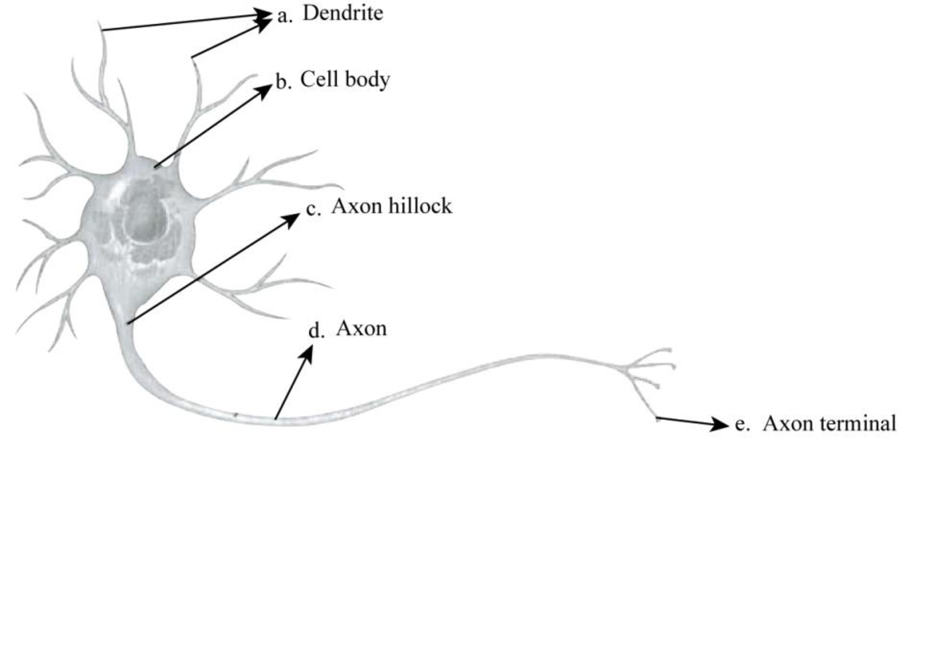
Fig.1: Structure of a neuron
Explanation of Solution
A neuron consists of a cell body, axon, dendrites, and terminal branches. The cell body is the largest part of the neuron; dendrites receive the signals, and then transmit them to axons, which further transfer them to the terminal branches. These signals originate from a region of cell body known as axon hillock. The synaptic terminals are present at the branching ends of the axons, which generally release neurotransmitters.
To determine: The direction of impulse transmission.
Introduction: An impulse refers to a signal transmitted along a nerve fiber. It is the way nerve cells communicate with each other.
Explanation of Solution
The direction of impulse transmission in a typical neuron is one direction. The dendrite receives the nerve impulse from other neurons, and a single axon transmits signals to other cells. The information is transmitted to another cell at the synapse. The synaptic terminals present at the ends of axons release neurotransmitters that relay signals to another neuron.
To determine: The event that occurs at Part e.
Introduction: The basic working unit of the brain is known as a neuron. It transmits information to other nerve cells, gland, or muscle cells.
Explanation of Solution
The branching ends of the axons are known as synaptic terminals. These terminals usually release neurotransmitters that relay signals to another neuron, a gland cell, or a muscle cell.
Synaptic terminals release neurotransmitters that transmit signals to another nerve cell, muscle cell, or gland cell.
Want to see more full solutions like this?
Chapter 48 Solutions
Study Guide for Campbell Biology


- Match the neuron structure with its correct function or description. 1. Structure that receives stimuli and carries impulses to the cell body Grey matter 2. Part of the neuron that releases a chemical transmitter across the synapse + Myelin sheath 3. Cells that protect, nourish, and defend neurons + White matter 4. Structure that contains the neuron's nucleus and is the site of the neuron's cell metabolism : Synapse 5. The gap between two myelin sheaths that allows faster conduction of action potential 6. Lipid, fatty insulating layer around some axons that protects the neurons and speeds the impulses along the neuron + Dendrite 7. Unmyelinated neurons in the CNS + Axon 8. Structure that transmits impulses away from the cell body to another neuron, the muscles, or glands Axon terminal 9. Myelinated neurons in the CNS Cell body 10. The gap or junction between two neurons or between a neuron and a muscle Node of Ranvier * Glial cellarrow_forwardDraw a neuron, label the following parts and give their functions: Cell body, nucleus, axon, myelin, schwann cell, nodes of Ranvier dendrites, synaptic knob, impulse,arrow_forwardDraw a pseudounipolar nerve synapsing with a unipolar neuron. Highlight the synapse and label the diagram.arrow_forward
- Describe the resting potential for neurons. How is this potential established? Describe the development of the action potential. How does this come about, and what is the function of this phenomenon? Include a diagram of the various stages of the action potential, showing the changes in voltage that occur throughout this phenomenonarrow_forwardGive the major function of each branch of the human nervous system Central Nervous System (CNS): Peripheral Nervous System (PNS): Give real life examples.arrow_forwardDescribe the organization of the nervous system. Distinguish between the functions of neurons and neuroglia. Describe the cell body of a neuron. Distinguish among the functions of sensory neurons, interneurons, and motor neurons. Summarize the functions of each of the types of neuroglia (the exam will not ask about satellite cells) and describe how the myelin sheath is formed around a peripheral nervous system neuron. Explain why nerve impulse propagation is compromised in patients with multiple sclerosis. Distinguish between the composition of white matter and gray matter. Summarize neuron communication from the moment of receptor stimulation to the response of an effector, such as a muscle fiber, and define neurotransmitter, resting membrane potential, and current. Define electrochemical gradients and the term “polarized”, and describe the electrochemical basis of the resting membrane potential including the function of the sodium-potassium pump in maintaining the resting membrane…arrow_forward
- View an electron micrograph of a cross-section of a myelinated nerve fiber image below. The axon contains microtubules and neurofilaments, bounded by a plasma membrane known as the axolemma. Outside the plasma membrane of the axon is the myelin sheath, which is composed of the tightly wrapped plasma membrane of a Schwann cell. What aspects of the cells in this image react with the stain that makes them the deep, dark, black color, such as the multiple layers that are the myelin sheath?arrow_forwardName the three primary structures of a neuron and explain the function of the nerve cell processes.arrow_forwardI. Identify the different structures of the neuron. A H. G E = A = F = В 3 G = C= H = D=arrow_forward
- Label the 4 major regions of the neuron and describe their functions.arrow_forwardLabel the type of neuroglia found in the CNS. Briefly describe each type.arrow_forwardDescribe how an action potential is generated in a neuron. Include channels that are open/closed during this process and be sure to include terms such as depolarization, threshold, hyperpolarization, and repolarization.arrow_forward

 Human Physiology: From Cells to Systems (MindTap ...BiologyISBN:9781285866932Author:Lauralee SherwoodPublisher:Cengage Learning